
Successful Treatments
Clinics
Doctors
Gout in the Knee: Symptoms and Diagnosis
 Published on 28 Sep, 2026
 Updated on 06 Oct, 2026

Explore Gout Joints, Stages and Tophi
- Home / Diseases & Ayurveda / Joint & Muscle Pain / Ayurvedic Treatment for Gout: Uric Acid, Joint Pain & Flare-Up Management / Understanding Gout Joints, Stages and Tophi / Gout in the Knee: Symptoms and Diagnosis
How Knee Gout Presents
A gout attack in the knee is characterized by its explosive, rapid onset. You may go to bed feeling completely normal and wake up just hours later in agonizing pain. Because the knee capsule is so large, a massive immune response causes it to rapidly fill with excess synovial fluid, a condition commonly called an effusion, or water on the knee.
The knee will appear dramatically swollen, often looking twice the size of your other knee, completely obscuring the natural bony outline of the kneecap. The skin stretched over the joint will turn red, tight, and shiny, radiating intense heat. The pain is severe and throbbing. Unlike a mild ache, gout pain is so hypersensitive that simply brushing a bedsheet against the knee can be intolerable. The immense swelling and severe pain will drastically limit your range of motion, often locking the leg in a slightly bent position.
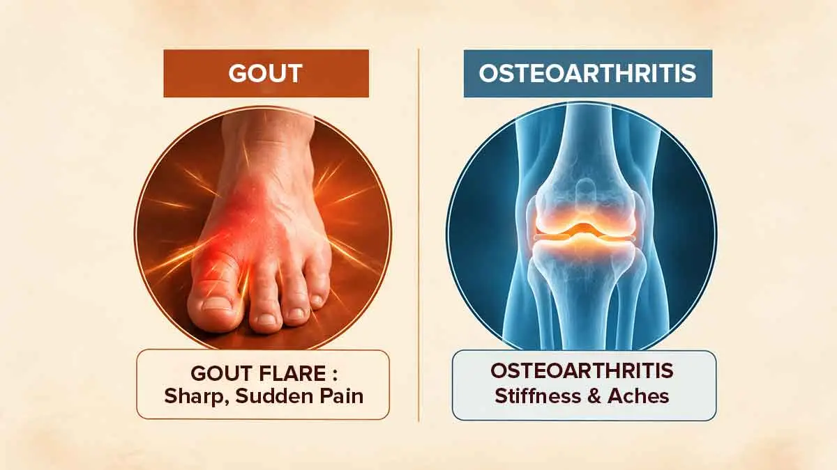
Gout Versus Injury And Osteoarthritis
Because the knee is so prone to injury and wear and tear, it is easy to confuse a mechanical problem with a metabolic one. However, the timeline of your symptoms is a massive clue.
Physical injuries such as a torn meniscus, a ruptured ACL, or a severe sprain usually have a distinct, memorable trigger. You twisted sharply, felt a “pop” during a run, or suffered a direct impact. Gout, conversely, appears entirely unprovoked, striking while you are at rest.
Osteoarthritis, the standard “wear-and-tear arthritis, develops incredibly slowly. It causes a dull, grinding ache and morning stiffness that gradually worsens over months and years as cartilage physically wears away. Gout is the exact opposite; it is an acute, sudden-onset firestorm that is at its peak within 24 hours in a joint that may have felt perfectly fine the day before.
 disease.webp)
Gout Versus Pseudogout
When it comes to the knee, gout has a nearly identical twin, pseudogout, medically known as Pyrophosphate deposition (CPPD) disease. Pseudogout is also caused by microscopic crystals forming in the joint space. However, instead of being made of uric acid, these crystals are made of calcium; crucially, while normal gout overwhelmingly prefers the big toe for its first attack, pseudogout’s absolute favourite target is the knee. To the naked eye, a pseudogout attack in the knee looks and feels exactly like a standard gout attack; it is red, violently swollen, hot, and intensely painful. You cannot tell the difference based on symptoms alone. Differentiating between the two requires a doctor to extract fluid from the knee and examine the specific chemical shape of the crystals under a microscope.
Why Infection Must Be Ruled Out
The most dangerous lookalike to knee gout is septic arthritis, a severe bacterial infection inside the joint capsule. The knee is highly susceptible to septic arthritis, and the symptoms are extreme swelling, redness, heat, and agonizing pain mimic a gout flare perfectly.
The distinction is a matter of life and death for your joint. While a gout flare will eventually subside on its own without permanent immediate damage, bacteria trapped inside the knee will begin rapidly eating away at your irreversible cartilage within a matter of days. Doctors must always consider the possibility of joint infection when presented with a hot, swollen knee, especially if the patient has a suppressed immune system, a recent cut or scrape near the leg, or a history of knee surgeries.
Examination And Joint-Fluid Testing
To cut through the confusion of these overlapping conditions, rheumatologists and emergency doctors rely on a definitive, gold standard procedure called arthrocentesis, or joint aspiration. The doctor will thoroughly numb the skin and insert a needle directly into the swollen knee capsule to draw out a sample of the excess synovial fluid. While this sounds daunting, patients often experience immense, immediate relief as the physical fluid pressure inside the tight knee is suddenly reduced. The extracted fluid is immediately sent to a lab and placed under a polarized light microscope. If the technician sees negatively birefringent, needle-shaped uric acid crystals, gout is definitively confirmed. If they see rhomboid-shaped calcium crystals, it is pseudogout. If they see a massive white blood cell count and bacteria, it is an infection.
Imaging And Blood Tests
While joint aspiration is the best diagnostic tool, doctors also utilize blood work and imaging to build a complete picture. Blood tests will be drawn to check your white blood cell count and inflammatory markers (like CRP and ESR) to help gauge the likelihood of a systemic infection. As with all gout flares, a blood uric acid test taken during the attack may be misleadingly normal, so it is used more for baseline tracking than acute diagnosis.
X-rays are routinely ordered to check for underlying osteoarthritis, calcification of the cartilage (a strong sign of pseudogout), or hidden fractures. More advanced imaging, such as a specialized musculoskeletal ultrasound, can easily peer inside the knee to detect the "double contour sign," a frosty, glowing layer of uric acid crystals coating the smooth surface of the knee's internal cartilage.
Attack Management Overview
Once an infection is strictly ruled out and gout is confirmed, the immediate goal is suppressing the immune system's violent reaction. Because the knee is a large, accessible joint, drawing the fluid out during diagnosis is often the first step in pain relief.
From there, doctors frequently rely on potent oral anti-inflammatory medications, such as high-dose NSAIDs (like Indomethacin) or Colchicine. However, for a severe knee flare, an intra-articular corticosteroid injection is often the most magical, fast-acting treatment. Because the doctor has already placed a needle into the knee to draw fluid for testing, they can seamlessly inject a powerful, localized dose of steroids directly into the inflamed space before removing the needle, often halting the attack entirely within 12 to 24 hours. At home, absolute rest, elevation of the entire leg, and cold therapy are required until the flare resolves.
When Urgent Care Is Needed
A hot, swollen knee should never be ignored, but certain accompanying symptoms transform the situation from a standard gout flare into a medical emergency.
You must seek urgent care or go directly to the emergency room if your swollen knee is accompanied by a high systemic fever, severe chills, shaking, or night sweats. If you notice red streaks creeping up your thigh or down your calf, or if the skin of your knee breaks open, these are massive red flags for spreading bacterial infection. Additionally, if the pain is so profoundly severe that you cannot bear a single ounce of weight on the leg without collapsing, do not wait for a routine clinic appointment; seek immediate emergency medical evaluation to rule out septic arthritis and protect your knee.
References
Gout: Diagnosis, Treatment, and Steps to Take
Find Jiva Ayurveda Clinics for Gout Treatment Near You
Consult Experienced Ayurveda Doctors for Gout














Our Happy Patients
-
 Sunita Malik - Knee Pain
 -
 Abhishek Mal - Diabetes
 -
 Vidit Aggarwal - Psoriasis
 -
 Shanti - Sleeping Disorder
 -
 Ranjana - Arthritis
 -
 Jyoti - Migraine
 -
 Renu Lamba - Diabetes
 -
 Kamla Singh - Bulging Disc
 -
 Rajesh Kumar - Psoriasis
 -
 Dhruv Dutta - Diabetes
 -
 Atharva - Respiratory Disease
 -
 Amey - Skin Problem
 -
 Asha - Joint Problem
 -
 Sanjeeta - Joint Pain
 -
 A B Mukherjee - Acidity
 -
 Deepak Sharma - Lower Back Pain
 -
 Vyjayanti - Pcod
 -
 Sunil Singh - Thyroid
 -
 Sarla Gupta - Post Surgery Challenges
 -
 Syed Masood Ahmed - Osteoarthritis & Bp
Diagnostic Marker
Blogs
- What is Gout? Causes and home treatment
- Are you suffering from Gout pain? Try these 5 natural home remedies
- List Of Foods To Avoid With Gout & Why
- Ayurvedic Approach to Gout Home Remedies in India
- सर्दियों में यूरिक एसिड और गाउट क्यों बढ़ते हैं? आयुर्वेद से जानिए दर्द से राहत का प्राकृतिक तरीका
- गाउट में बार-बार दवाइयाँ लेने से किडनी पर क्या असर होता है? और आयुर्वेद कैसे राहत देता है
- Gout के मरीज़ को किन चीजों से तुरंत सावधान हो जाना चाहिए?
- Uric Acid Test Normal आया पर Joint Pain बना हुआ है — Pseudo-Gout हो सकता है
- Gout Attack बार-बार क्यों लौटता है जबकि दवा चालू है? असली Trigger समझिए
- क्या सिर्फ खान-पान से uric acid control हो सकता है? पूरा सच जानिए
Home Remedies
Related Disease
- Ayurvedic Treatment for Ankylosing Spondylitis: Pain, Symptoms, Causes & Care
- Ayurvedic Treatment for Cervical Spondylosis: Neck Pain, Stiffness & Management
- Ayurvedic Treatment for Arthritis: Joint Pain Relief & Long-Term Management
- Ayurvedic Treatment for Osteoarthritis: Knee Pain, Symptoms and Management
- Ayurvedic Treatment for Gout: Uric Acid, Joint Pain & Flare-Up Management
- Ayurvedic Treatment for Sciatica: Managing Nerve Pain & Leg Discomfort
- Ayurvedic Treatment for Rheumatoid Arthritis: Joint Swelling, Symptoms & Care
- Ayurvedic Treatment for Frozen Shoulder: Pain, Symptoms, Causes & Care
- Ayurvedic Treatment for Deep Vein Thrombosis (DVT): Leg Swelling, Risks and Treatment
- Ayurvedic Treatment for Achilles Tendon Disorders: Pain, Symptoms, Causes & Care
- Ayurvedic Treatment for Rickets: Symptoms, Causes & Management
- Ayurvedic Treatment for Ganglion Cyst: Symptoms, Causes & Management
- Ayurvedic Treatment for Osteoporosis: Pain, Symptoms, Causes & Care
- Ayurvedic Treatment for AVN Avascular Necrosis: Pain, Symptoms, Causes & Care
- Ayurvedic Treatment for Osteomyelitis: Symptoms, Causes & Care Options
- Ayurvedic Treatment for Cerebral Palsy: Symptoms, Causes & Supportive Care
Latest Blogs
- Late Dinner Plus Poor Sleep: Why Ayurveda Looks At The Pattern, Not One Habit
- परिवार के साथ बैठकर खाना खाने से Portion और Eating Speed पर क्या फर्क पड़ सकता है?
- कब्ज़ में इंडियन-स्टाइल टॉयलेट बेहतर है? टॉयलेट पोस्चर के बारे में क्या पता है
- दूध, दही और लस्सी से IBS के लक्षण कब बढ़ सकते हैं?
- कभी कब्ज़, कभी दस्त? बार-बार बदलती पेट की आदतों को कैसे समझें
- क्या आयरन, कैल्शियम या कुछ दवाओं से कब्ज़ हो सकती है? डॉक्टर से कब बात करें
- पीरियड्स के दौरान IBS के लक्षण कुछ लोगों में क्यों बढ़ सकते हैं?
- व्रत या उपवास में कब्ज़ बढ़ जाती है? नवरात्रि में किन बातों का ध्यान रखें
- सफ़र में कब्ज़ क्यों हो जाती है? यात्रा से पहले क्या तैयारी करें
- खूब पानी पीने के बाद भी कब्ज़ क्यों बनी रह सकती है?
- बुज़ुर्ग माता-पिता को कब्ज़ रहती है? परिवार किन बातों का ध्यान रखे
- क्या खूनी बवासीर में ऑपरेशन ही एकमात्र विकल्प है? इलाज के विकल्प समझें
- शौच में खून देखकर घबरा गए? जानिए पहले क्या समझें और क्या करें
- रोटी, चावल और दही: कब्ज़ होने पर खाने में क्या बदलाव मदद कर सकते हैं?
- रोज़ बाइक से ऑफिस जाते हैं और बवासीर की सूजन परेशान करती है? जानिए सफ़र का असर
- IBS में दवा से राहत मिलने के बाद भी लक्षण दोबारा क्यों हो सकते हैं?
- IBS में कौन-सा खाना परेशानी बढ़ाता है? फ़ूड डायरी कैसे मदद कर सकती है
- मानसून में पेट की परेशानी बढ़ती लगती है? IBS और मौसम के संबंध को समझें
- बवासीर में सिट्ज़ बाथ कैसे करें? फायदे, सावधानियाँ और कब बचें
- बवासीर के मस्से बाहर निकलते हैं? ग्रेड 1 से 4 का मतलब समझें
Ayurvedic Doctor In Top Cities
- Ayurvedic Doctors in Bangalore
- Ayurvedic Doctors in Pune
- Ayurvedic Doctors in Delhi
- Ayurvedic Doctors in Hyderabad
- Ayurvedic Doctors in Indore
- Ayurvedic Doctors in Mumbai
- Ayurvedic Doctors in Lucknow
- Ayurvedic Doctors in Kolkata
- Ayurvedic Doctors in Patna
- Ayurvedic Doctors in Vadodara
- Ayurvedic Doctors in Ahmedabad
- Ayurvedic Doctors in Chandigarh
- Ayurvedic Doctors in Gurugaon
- Ayurvedic Doctors in Jaipur
- Ayurvedic Doctors in Kanpur
- Ayurvedic Doctors in Noida
- Ayurvedic Doctors in Ranchi
- Ayurvedic Doctors in Bhopal
- Ayurvedic Doctors in Ludhiana
- Ayurvedic Doctors in Dehradun